Identify All Indicated Parts of the Nerve Section
Region of the cell body from which the axon originates a. When you look at the spinal cord cross section it almost looks like a taffy candy that has a butterfly right in the middle.

Schematic Representation Showing The Structures Attaching To The Coracoid On A Right Shoulder The Anterior Cora Medical Anatomy Anatomy And Physiology Anatomy
Identify the nerve highlighted in the image.

. Select the answer in which these alphabets have been correctly matched with the parts which they include. Collection of nerve cell bodies found outside the CNS. Receptive region of a neuron c.
ASTROCYTES HELPS REGULATE THE EXTRACELLULAR COMPOSITION OF BRAIN FLUID. Identify the muscle indicated by B Rhomboid minor Match the following description of muscles that move the head and trunk with its appropriate name. Sketch the indicated view of a nerve fiber axon.
Cell body 2Neuronal membrane3Dendrites 4. A nerve is an enclosed cable-like bundle of axons the projections of neurons in the peripheral nervous system PNS. Insulates the nerve fibers d.
Identify the components of nerve shown in the photomicrograph longitudinal section. Gyrus and sulci singular. It produces all the proteins for the dendritesaxons and synaptic terminals and contains specialized organelles such asthe mitochondria Golgi apparatus.
EPENDYMAL CELLS SECRETES CEREBROSPINAL FLUID HELPS MOVE CEREBROSPINAL FLUID IN BRAIN AND SPINAL CORD. Neurogl ia neurotransmitters h. Different parts of the spinal cord and between the cord and the brain.
Nerve fiber cross section with Schwann. The brain and spinal cord collectively. Nerve Structure Anatomy Nerves are the organs that make up the peripheral nervous system PNS.
ANATOMY OF THE SPINAL CORD- CROSS SECTION PRE-LAB. Identify the cranial nerve which controls all but one of the muscles of the palate pharynx and the intrinsic muscles of the larynx. Central nervous system efferent neuron d.
Human Anatomy Physiology Laboratory Manual. Site of the nucleus and is the most. Match each statement with a response chosen from the key.
A nerve provides a structured pathway that supports the electrochemical nerve impulses transmitted along each of the axons. Neuron that conducts impulses away from the CNS to muscles and glands. Inside the nerves groups of neurons nerve cells are organized into bundles called fascicles fasciculi.
They are each also divided into subparts or regions for simplified localization of structures for example. INCLUDE ANDD LABEL THE FOLLOWING STRUCTURES ON. MAY BE INVOLVED IN THE TRANSPORT OF SUBSTANCES WITHIN THE NEURON.
It is connected to the two fallopian tubes on its superior end and to the vagina via cervix on the inferior end. Microscope image of full optic nerve cross section from 66 year old AD sample. Histology of Nervous Tissue Flashcards.
It has many nerve tracts groups of axons traveling together moving up ascending and down descending its length. It is a branch of the trigeminal nerve the fifth cranial nerve which serves both a. B- Osteocytes C- Haversian system.
Neuron that conducts impulses toward the CNS from the body periphery. Match the following anatomical terms column B with the appropriate description or function column A. In the central nervous system the analogous structures are known as tracts.
Axon hillock E 4. DRAW A TYPICAL MULTIPOLAR NEURON. IMPULSE GENERATOR AND TRANSMITTER.
A very large fast-adapting tactile receptor that is composed of a single dendrite enclosed by concentric layers of collagen is a. Sulcus of the cerebrum. Cell body The soma is the factory of the neuron.
Composite muscle forming part of the deep layer of intrinsic back muscles located along the back from thoracic region to head. Read the introduction to the lab and answer the following questions. Cerebrospinal fluid enters the central canal of the spinal cord from the.
Parts of the Nerve Cell and Their Functions Silvia Helena Cardoso PhD 1. A tactile receptor composed of highly coiled dendrites that are surrounded by modified Schwann cells and a fibrous capsule is a. Dark sections indicate bundles of axons separated by the lighter connective tissueA magnified section of axons is provided characterized by their circular shape and lighter center surrounded by dark myelin sheaths.
The midsagittal section of the brain shows the three major parts of the brain which are the cerebrum cerebellum and brainstemThese brain parts are marked with visible gross features like the gyri singular. Axon terminal D 3. ESSENTIALLY ROUGH ENDOPLASMIC RETICULUM IMPORTANT METABOLICALLY.
The uterus is a hollow muscular pear-shaped organ located posterior and superior to the urinary bladder. Column A Column B C 1. They serve as information pipelines that allow the brain and spinal cord to communicate with other tissues and organs.
Label any structures observed and indicate the magnification. Tasked with carrying important information to the central nervous system CNS the maxillary nerve runs from upper gingiva the upper set of teeth along the surface of the middle of the face through the palate and nasal cavity before terminating in the upper lip and cheek. The vagina is an elastic muscular tube that connects the cervix of the uterus to the exterior of the body.
This area is made up of all the nerve fibers that direct the reflex actions and convey the impulses that go back and forth to the brain.

The Entire Neuro Chapter From Page 318 To 363 In One Long Scroll Cool Tasman W Jaeger E A Wills Eye Hospita Optometry Education Optometry Medical Knowledge

Eye Anatomy Diagram Illustrations 41 New Ideas Eye Anatomy Eye Anatomy Diagram Human Body Anatomy

Health Secret Hidden In Your Fingers Su Jok Therapy Can Prevent Serious Diseases Care Body Hai Acupressure Points Acupressure Acupressure Points In Hand
Basic Structure And Function Of The Nervous System Anatomy And Physiology

The Nervous System

Histology Of The Peripheral Nerves And Light Microscopy Nysora Nysora

Histonano Com Books Junqueira S 20basic 20histology 20pdf 20whole 20book 22 20the 20fema Anatomy And Physiology Reproductive System Female Reproductive System

The Nervous System

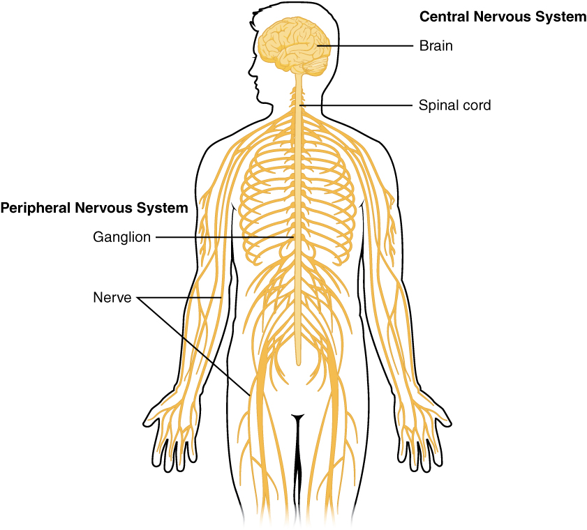
Parts Of The Nervous System Introduction To Psychology

Labelled Diagram Of The Ear Ear Diagram Ear Anatomy Human Ear Diagram

Histology Of The Peripheral Nerves And Light Microscopy Nysora Nysora

Pin On Spinal Columan Anatomy

Nerves Of The Upper Limb An Overview Sciencedirect Topics

Histology Of The Peripheral Nerves And Light Microscopy Nysora Nysora

Basic Structure And Function Of The Nervous System Anatomy And Physiology I

Spinal Nerve Definition Function Diagram Number Facts Britannica

Vagus Nerve Vagus Nerve Stimulator Reflexology

Parts Of The Nervous System Introduction To Psychology
Do You Suffer From Overactive Upper Trapezius Or Traps Lowerbackpain Brachial Plexus Products Thoracic


Comments
Post a Comment